 Saprolegnia
spp.
 Saprolegnia
spp.
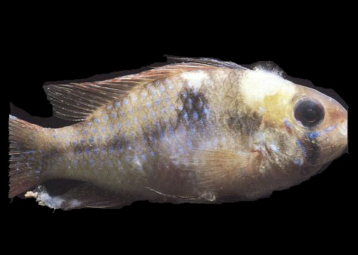
Saprolegnia spp. are aquatic fungi that occasionally are found infecting skin wounds of tilapia in Hawaii. Although tilapia are rather resistant to fungal infections, wounds that occur during handling or with crowded conditions may be colonized by the fungi. Saprolegnia spp. proliferates in organic matter poor water quality and organic build-up on the tank bottom will enhance the abundance of this fungus. Although Saprolegnia spp. are a frequent problem to eggs of other species of fish, this problem is not recognized in tilapia (probably because these fish are mouth brooders).
 Presumptive
diagnosis of Saprolegnia sp. infection is by demonstration of the cottony
growth of the fungus protruding from wounds on the fish. Microscopic examination
of wet mounts is needed to show the presence of the fungal hyphae and rule
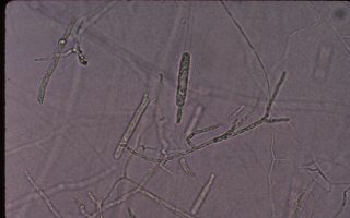
out Epistylis sp. Microscopically (100X to 400X) Saprolegnia spp. and other
types of aquatic fungi appear as branching mass of þroot-likeþ
filaments (e.g. the hyphae). The filaments of the fungus are clear and about
5 þm in diameter.
Saprolegnia spp. develop near the end of specialized hyphae the reproductive structure a club-shaped sporangia. The sporangia is important for identification. Motile zoospores are produced and released from the sporangia. These zoospores are the means that the fungus spreads. Zoospores settle on a suitable substrate (e.g. open sore or in organic matter on the bottom) and produce the vegetative hyphae of the fungus.
Control of Saprolegnia spp. should be aimed and timely removal of organic matter, avoidance of wounding of fish and maintenance of healthy water quality conditions. Saprolegnia spp. infection have been reported to respond to treatment with malachite green. If the fungus has invaded into the host tissue, the treatment will not be destroy the fungus in these locations. Malachite green has not received FDA approval for use with fish and this chemical should not be used with tilapia cultured in Hawaii.
See also: